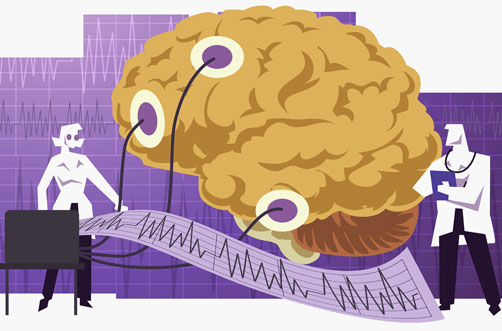
Electroencephalogram of the brain - principle of action and methods of application
If a person, who is in a state of mental and physical rest, applies electrodes to the head and through an amplifier connects them to a recording device, then you can catch electrical vibrations… These vibrations originate in the cerebral cortex and are associated with special nervous activity. They are also recorded directly from the brain when the skull is opened during surgery.
The presence of rhythmic, spontaneously occurring electrical oscillations in the brain was established in 1875 by the Russian physiologist V. Ya. Danilevsky and the English scientist Richard Cato, independently of each other, experimenting on animals with an open skull.
It was subsequently shown that it was possible to record electrical currents of the brain through the skin and bones of the intact skull. This served as the basis for the transition to the study of these phenomena in humans.
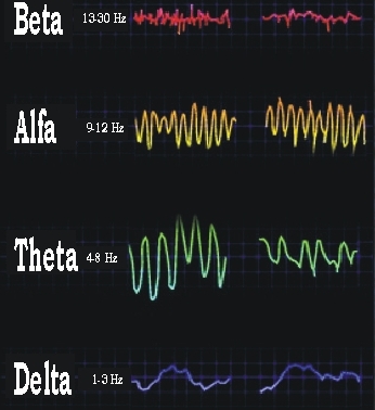
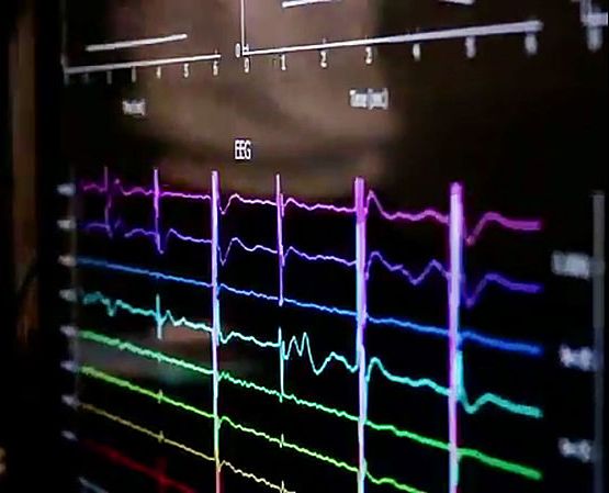
The most interesting feature of the electrical vibrations of the human brain is their characteristic, almost regular rhythm with a frequency of about 10 Hz - these are the so-called alpha waves.In their background, more frequent oscillations are visible - beta waves at 13 - 30 Hz and gamma waves at 60 - 150 Hz and above. Slower oscillations are also observed - waves of 1 - 3 - 7 Hz.
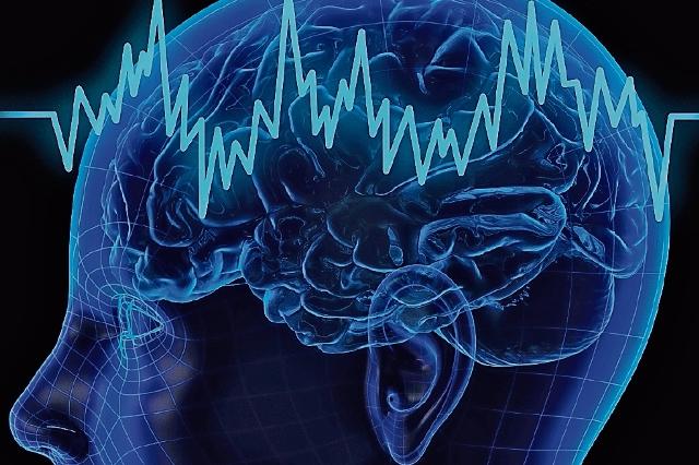
The electrical waveform of the brain is called an electroencephalogram, and the branch of electrophysiology that studies the patterns of electrical activity in the brain is called electroencephalography (EEG). Electroencephalograms lend themselves to Fourier mathematical analysis.
Electroencephalography is of great importance for theoretical studies of brain activity, as well as for practical purposes of diagnosing brain diseases.
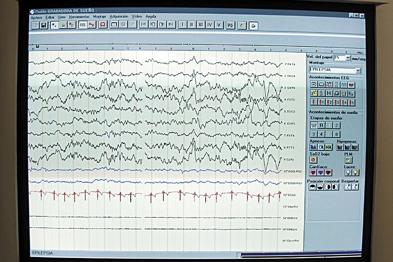
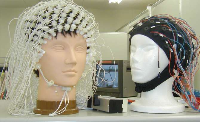
To protect the object from external electromagnetic fields, it is placed in a shielded room. Sources of errors in electroencephalogram acquisition: skin and muscle potential, electrocardiogram, arterial pulsation, electrode movement, eyelid and eye movement, and amplifier noise.
The best electroencephalogram is obtained from a person at complete rest: a person is sitting or lying down better (but not sleeping) in a screened soundproof dark room in a comfortable position, isolated from external stimuli and at complete rest.
This circumstance is very important. Often in people who come to the study for the first time, it is difficult to register an electroencephalogram due to their vigilance and fear of an unusual environment.
People differ from each other in their inherent EEG characteristics. In some it is very easy to detect the correct rhythm of alpha waves, in others it is not recorded at all.
Electroencephalograms also differ in shape, amplitude, duration, regularity of alpha waves, as well as in the location, number and intensity of other waves - beta, delta and gamma.
It is interesting to note the surprising constancy of the basic features of the human electroencephalogram, established by repeated studies over many months.
It is usually possible to know in advance how soon a regular electroencephalogram will be established in a well-studied subject and what his characteristic features are. However, along with the great constancy of the distinctive features of the individual electroencephalogram of a healthy person, there is also a great physiological variability of it, even during the same day.
An indispensable condition for obtaining a regular electroencephalogram of a person is the exceptional rest of the waking brain. It is understandable how difficult it can be to achieve this in an energetic state, turning off brain activity.
By observing for hours, day after day, the electrical vibrations occurring in a person's cerebral cortex, one can see that the brain is often like a mirror, reflecting what one is doing at the moment.
Sometimes the regular rhythms of the brain suddenly disappear on their own, or high-frequency oscillations appear, or special muscle currents appear. It means that the person thought about something, made some movement, imagined something. The variability of the electroencephalogram reflects fluctuations in the excitability of the central nervous system.
If you ask a person to perform some mental work, for example, solving a problem representing a difficult situation, then you can observe the disappearance of the regular rhythm of alpha waves and the appearance of high-frequency oscillations. During intense mental work, alpha waves are replaced by high-frequency discharges of 500-1000 Hz, lasting throughout the duration of mental activity, after the termination of which alpha waves are restored.
High-frequency oscillations associated with mental activity can last for a long time. In a student who usually establishes a normal brain rhythm, it becomes difficult to record an EEG — only high-frequency oscillations are observed. It turned out that he was busy preparing for the exams on his days free from experiments.
In another subject who had a normally regular electroencephalogram with surprising ease, only high-frequency oscillations were observed once. It turned out that he had been drawing for two hours before the experiment.
In general, a normal rhythm of alpha waves is characteristic of the human brain in a calm state, and high-frequency oscillations, beta and gamma waves, are associated with its activity.
The rhythmic activity of the brain, in addition to the motor area, begins in a person only a month after birth; it develops, apparently, simultaneously with cortical activity as the child begins to recognize and grasp objects.
Since at this age it is different from that of adults, the electroencephalogram gradually changes, only by 11-12 years of age it approaches the norm for an adult.The rhythmic activity of the brain continues in sleep, but changes, becomes more simplified and smooth, slow vibrations appear.
It is interesting to note that the rhythm of the sleeper's brain is not disturbed, for example, by the noise of a car from the next room or the sound of a horn from the street, but if a sound is heard in the room, for example, the rustling of paper, associated with the fact that there is someone in the room.the sleeper's brain changes. This is due to the presence of «observation points of the brain», which are awake during a person's sleep.
With the help of the electroencephalographic method, it is possible to objectively observe and record these complex changes in brain activity associated with a certain subjective sensation.
In brain disease, waves of a special shape and duration appear. In brain tumors, slow waves with a frequency of 1-3 Hz appear, which he calls delta waves. Delta waves are recorded when picked up from the point on the skull directly above the tumor, while when picked up from other areas of the brain not picked up by the tumor, normal waves are recorded. The appearance of delta waves in the part of the brain affected by the tumor is determined by the degeneration of the cortex in this place.
In this way, the electroencephalogram helps to recognize the presence of a tumor and its exact location. Delta waves in the electroencephalogram are also found in other pathological conditions of the brain.
In some traumas: pathological delta waves are observed in the electroencephalogram many years after a head injury.
The rhythms of the human brain change or disappear completely with a loss of consciousness caused by various reasons, they change with a lack of oxygen. Thus, in experiments studying the effect of breathing in a mixture of air with a reduced percentage of oxygen, which caused a loss of consciousness , groups of spike-like waves, exceptional in voltage, are recorded, as if the brain has lost some kind of brake.
The same spasmodic slow waves have been recorded in people unconscious from concussion immediately after a head injury. In some brain diseases, high-frequency potentials are registered (for example, in schizophrenia) or in the alternation of slow wave and wave (in epilepsy).
The method of electroencephalography is essential for the diagnosis and study of brain diseases. As for the theoretical importance, electroencephalography, allowing to register the state of excitability of the cerebral cortex, opens access to the direct study of the processes of excitation and inhibition in the human brain, the ratio of which is considered the main mechanisms of nervous activity.